
A better imaging experience for your patients.
Dramatically improve your patient breast screening compliance rates with the Vera Scan by Koning.
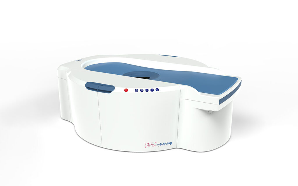
The Vera Scan by Koning
FDA approved for Diagnostic Breast Screening
The Vera Scan by Koning is the only FDA-cleared dedicated breast CT system. Unlike a mammogram, it creates a full isotropic 3D image of each breast.
The Technology
Dedicated Breast CT (DBCT) - a new standard of testing.
Clinical Evidence
Peer-reviewed research supports Dedicated Breast CT.
Multiple institutions have studied Dedicated Breast CT and published findings in leading radiology journals. The evidence demonstrates comparable or superior lesion detection, improved patient tolerance, and strong radiologist confidence in image quality. The following studies are ordered by publish year.
IMPORTANT NOTICE - October 2025: California Department of Public Health (CDPH) recognizes Dedicated Breast CT systems under the state Mammography Program establishing DBCT as an accepted clinical modality in California. View CDPH Mammography Facility Program
Sensitivity Comparison
DBCT vs. Mammography - Yang 2024
Pooled sensitivity. DBCT sensitivity is 19% higher in relative terms (0.92 vs. 0.77).
Diagnostic performance of dedicated breast CT for detection of breast cancer
Dedicated breast CT demonstrated high sensitivity and specificity for breast cancer detection, with radiologists reporting high confidence in image quality and lesion characterization.
Barriers to mammography screening among minority women in the United States: a systematic review of 17 studies
Pain and embarrassment associated with screening mammography were the most consistently reported barriers across minority populations. This finding is directly relevant to the Southern California patient population, where Filipino, Vietnamese, Chinese, Korean, and Hispanic women represent a disproportionately large share of the dense breast tissue population with documented compliance gaps.
Dedicated breast CT versus digital mammography: comparison of diagnostic accuracy
Dedicated breast CT showed non-inferior diagnostic accuracy compared to digital mammography, with significantly higher patient comfort scores and willingness to return for follow-up imaging.
Review of clinical studies and first clinical experiences with a commercially available cone-beam breast CT in Europe
Clinical experience with cone-beam breast CT demonstrated excellent visualization of breast parenchyma and lesions, with particular advantages in dense breast tissue and post-surgical evaluation.
The effect of mammography pain on repeat participation in breast cancer screening: a systematic review
In the most robust studies, 25 to 46% of women who did not re-attend mammography cited pain as the primary reason. Women who previously experienced mammography pain were statistically more likely to fail to re-attend screening (RR 1.34; 95% CI: 0.94 to 1.91). The authors concluded that sufficient evidence exists to confirm that painful mammography contributes to non-re-attendance, and called for effective pain-reducing interventions. Published in The Breast, 2013.
Study summaries are provided for informational purposes. Refer to original publications for full methodology and results.
Actuarial Sensitivity Analysis
Patient Compliance Projections
In the absence of large-scale published compliance outcome data specific to DBCT, expected given the technology's early deployment, the projections below are presented as a structured sensitivity analysis.
Below you will find three compliance scenarios based on the pain-compliance relationship documented in the Whelehan 2013 systematic review, with operator case study data from deployed DBCT sites serving as the real-world reference for the Target scenario. We fully expect to reach the Target scenario with the removal of traditional breast compression; dramatically improving patient compliance rates.
20% relative improvement over current mammography compliance rates
Lower bound of Whelehan pain-attribution data; assumes partial but not complete elimination of pain-driven non-attendance
Modest increase in screening volume; material improvement in Stage 0-1 detection rates
50% relative improvement
Mid-range of Whelehan data; consistent with published direction of pain-compliance relationship across multiple study designs
Material increase in screening volume; significant earlier-stage detection
Up to 100% relative improvement
Consistent with operator case study data from deployed DBCT sites; upper bound assumes near-complete elimination of compression as compliance barrier
Highest screening volume; maximum downstream Stage 0-1 benefit
Source: Whelehan 2013 systematic review (pain-compliance relationship); DBCT operator case study data (high scenario).
Diagnostic Screening
Gentle only accepts "Diagnostic Screening" referrals at this time. These patient scenarios qualify.
Dense breast tissue
Patients with heterogeneously or extremely dense breasts (BI-RADS C or D) where mammographic sensitivity is reduced.
Diagnostic workup
Patients with a palpable abnormality, nipple discharge, or asymmetry requiring further characterization beyond screening mammography.
High-risk screening
Patients with elevated lifetime risk due to family history, genetic mutation, or prior biopsy findings who benefit from supplemental imaging.
Implant evaluation
The Vera Scan provides a complete 3D volumetric assessment of both implant integrity and surrounding breast tissue — delivering implant screening and cancer screening in a single visit.
Post-treatment follow-up
Patients following lumpectomy, radiation, or other breast-conserving treatment where scar tissue complicates mammographic interpretation.
Patient anxiety or pain
Patients who have avoided or delayed screening (non-compliance) due to pain, anxiety, or prior negative experiences with compression-based imaging.
Referring Patients
Simple, straightforward referrals.
Download the referral form
Use our standardized referral form to order the Vera Scan. Return the completed form to our office. We'll confirm receipt, and contact your patient directly for scheduling.
Return the form to us with prior imaging
If you have any relevant patient prior imaging history please provide it, or use the notes section of the form to provide contact information for who we can contact to obtain prior images.
Receive the report
A board-certified radiologist with specialized CT breast imaging expertise will review the study. A detailed report will be sent to your office within 1 to 2 business days of your patient's Scan.
The referral form will be available when we are accepting patients.
Pricing & Payment
Simple, transparent pricing.
Gentle Breast Imaging will operate on a self-pay model until insurance companies offer this benefit. Your patient will have payment options, but this service is not reimbursed by Payers yet. We will roll out reimbursements as they are approved by payers and keep you informed.
Flexible payment
Payment options
We get it. Paying cash for care can be hard; especially when your patient is already paying for insurance. That is why we offer three ways to spread the expense so their health does not have to wait.
Simple payment plan - 50/50
Pay 50% when scheduling the appointment, and the remaining 50% on the date of service. No interest. No fees. Patients can spread the expense over pay dates or credit card clearing dates.
HSA and FSA payments
Health Savings Accounts (HSA) and Flexible Spending Accounts (FSA) are funded with pre-tax dollars, making your patient's pay go further toward this out-of-pocket expense.
Credit and debit cards
We accept all major credit cards and debit cards. Pay in full at the time of the appointment and earn those points or miles.
Partner With Us
Get in touch with our team.
Whether you're ready to refer patients, want to learn more about the technology, or are interested in a contracting conversation, we'd love to hear from you.